


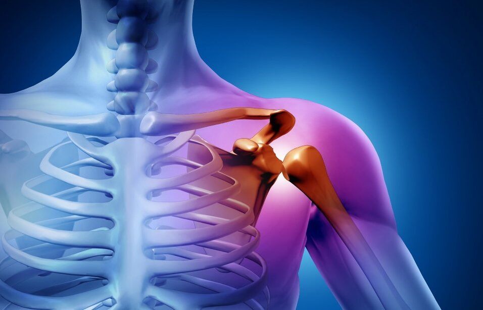
Shoulder osteoarthritis (arthropathy) is a chronic disease in which an irreversible degenerative dystrophic process occurs in joint tissue. The pathology disrupts the normal function of the limb. The range of motion of the shoulder is gradually reduced to complete immobility. Shoulder osteoarthritis can cause severe pain and reduce quality of life. Disability occurs without treatment.
In order to stop the joint destruction process and maintain the mobility of the shoulder joint, it is necessary to contact an orthopaedic traumatologist after the first symptoms.
Causes of shoulder osteoarthritis
The disease has multiple etiologies. The development of shoulder deforming arthropathy may be related to a variety of factors:
- Professional sports or high-intensity training.
- endocrine diseases.
- hormone imbalance.
- Congenital disorders of the development of the musculoskeletal system.
- Genetic predisposition, etc.
In most cases, secondary arthropathy is diagnosed: pathology that develops after exposure to a joint of one factor or another. Primary or idiopathic forms of the disease are rarely documented. In this case, it is impossible to determine the exact cause of tissue degeneration.
Frozen shoulder symptoms
Changes in cartilage and bone tissue begin long before the first signs of arthropathy. Joint structures have great potential for self-healing, so lesions are rarely diagnosed at a young age when all metabolic processes are very active. As the body ages, the recovery process gives way to degeneration. The first signs of destruction may appear after 40-50 years, and with dysmorphic disease, patients notice changes as early as 16-18 years of age.
Frozen shoulder symptoms:
- Cracked joints during exercise.
- Pain, especially severe after exercise.
- Stiffness of movement, manifested after sleep or prolonged rest.
- The pain gets worse when the weather changes.
Degree of joint disease
The clinical classification defines three degrees of arthropathy of the shoulder joint:
- 1 degree. The patient complained of slight tightening during exercise. No pain syndrome. Discomfort when hands are placed in extreme positions.
- 2 degrees. Pain occurs when the limb is raised above the shoulder. Range of motion is reduced. After strenuous exercise, patients experience pain even at rest.
- 3 degrees. The range of motion of the joints is severely limited. Pain syndrome is almost constant.
Diagnosis of shoulder osteoarthritis
Doctors need not only to make a correct diagnosis, but also to determine the pathological cause. Treating the underlying disease can significantly improve a patient's health and slow cartilage degradation.
Manual inspection
The first stage of diagnosis is consultation with an orthopaedic trauma specialist. Doctors check the affected joint for swelling and severe deformity. From the point of view of the development of arthropathy, the muscles can partially atrophy - this can be seen with the naked eye.
With manual examinations, doctors assess the function of the joints based on several criteria:
- Able to make voluntary hand movements.
- Thickening of the articular margins (large osteophytes on palpation).
- A creaking, "clicking" sound that can be heard or felt in the hand during shoulder movement.
- Stuck the joint in the presence of free cartilage bodies.
- Pathological movement of the shoulder.
radiography
To detect signs of shoulder arthropathy, radiographs are performed in two projections, which allow you to assess how narrow the joint space is, the condition of the bone surface, the size and number of osteophytes, the presence of fluid, and inflammation of the surrounding tissue.
Ultrasound (ultrasound)
A non-invasive method that allows you to examine the joints of pregnant women and young children. From the sonogram, the doctor determines the thickness of the cartilage, the condition of the synovium. This method provides a good visualization of osteophytes, swollen lymph nodes in the periarticular space.
Magnetic Resonance Imaging (MRI)
An MRI machine takes pictures of serial sections. The image clearly shows not only the joint, but also the adjacent tissue. Magnetic resonance imaging is by far one of the most informative methods in the diagnosis of arthropathy.
lab testing
As part of the comprehensive exam, they appoint:
- General blood analysis. From the results, the doctor can judge the presence and severity of the inflammatory process. The analysis also helps assess overall health.
- Urinalysis. Kidney disease often leads to secondary joint deformities. Analysis is necessary for accurate diagnosis.
- blood chemistry. These data help determine the cause of inflammation. Biochemical analyses are also performed to monitor complications and side effects during treatment.
Treatment of Shoulder Osteoarthritis
Treatment is long and difficult. The course of treatment includes medication, wellness procedures, a set of special exercises for shoulder arthropathy. In difficult cases, surgical intervention is required.
medical treatement
Medications and doses are selected individually. A doctor may prescribe:
- Non-steroidal anti-inflammatory drugs (NSAIDs). Medications can reduce inflammation and pain.
- Glucocorticoid preparations. Hormone-based approaches have a stronger effect on pain focus. The drug not only relieves the patient's condition, but also reduces inflammation, exhibits antihistamine and immunosuppressive properties. Glucocorticoids are used when non-steroidal anti-inflammatory drugs are ineffective.
- painkiller. This group of drugs is used to treat severe pain syndromes. Depending on the severity of your symptoms, your doctor will choose a non-narcotic or narcotic (rarely) pain reliever.
- chondroprotective agent. The active ingredient of the drug is involved in the formation of new cartilage tissue. Regeneration of diseased joints is accelerated and nutrition is improved. Chondroprotective agents have a cumulative effect and have proven themselves in the treatment of arthropathy of varying severity.
Some drugs are injected directly into the joint cavity. For example, blockers have better pain relief than taking the drug in tablet form.
physiotherapy
The lessons are carried out after the aggravation has been eliminated. Physiotherapy, as part of a complex therapy, helps to improve the transport of medicines to the affected joint, reduce swelling and reduce pain.
For the treatment of joint diseases:
- electrophoresis.
- Acoustic swimming.
- Shockwave therapy.
Physiotherapy can be combined with massage, kinesiotherapy, therapeutic baths. A set of procedures is best performed in a specialized clinic. A doctor will develop a treatment plan based on a particular patient's condition.
physiotherapy
Moderate physical activity is important to slow down the degenerative process. Exercise therapy for shoulder joint disease is best started at a medical center under the supervision of a doctor. Experts will choose exercises that teach them how to perform correctly and distribute loads so that they don't worsen the disease. Gymnastics usually includes warm-ups, stretching, and strength training. Do exercise at least 3 times a week.
After the specialist's course, the patient can perform treatment exercises for shoulder arthropathy at home.
Operation
The surgery is performed in the case of 3rd degree arthropathy, when the disease no longer prevents the patient from moving normally, causes severe pain, and prescribed treatments do not help.
There are several methods of surgical treatment:
- prick. A long needle is inserted into the joint cavity to pump the accumulated fluid out. Puncture reduces pressure, reduces swelling, and increases joint mobility. The procedure is minimally invasive, so it is performed on an outpatient basis. The material obtained during the puncture process is sent for research to determine the source of infection or other indicators.
- Arthroscopy. With the help of microsurgical instruments, doctors examine the joint cavity, remove scar tissue, and suture the tendons of the rotator cuff or joint capsule if they are damaged. Several puncture points were left on the skin. The patient recovered quickly.
- Built-in prosthesis. An endoprosthesis allows you to completely free yourself from chronic pain and restore arm mobility. After surgery, long-term (3 to 6 months) rehabilitation is required.